Opn1mw
Medium-wave-sensitive opsin 1 (Green cone photoreceptor pigment) (Green-sensitive opsin) (M opsin) (Medium wavelength-sensitive cone opsin)
Spatial tier: REGION_BIASED · Class: GPCR · Top region: TMd (Tuberomammillary nucleus, dorsal part) · Positive regions: 32/554
Function in TMd: evidence, prediction, innovation
- Gene function
- OPN1MW is medium-wavelength (green/M) cone opsin, the light-sensing GPCR of cone photoreceptors. Bound 11-cis-retinal isomerises on photon absorption, activating transducin and the phosphodiesterase cascade that closes CNG channels; in mice M-opsin is expressed in a dorsoventral gradient across the cone mosaic.
- Region function
- The dorsal tuberomammillary nucleus contains the brain's only histaminergic neurons; they project diffusely to cortex, thalamus and hypothalamus and promote wakefulness, arousal and attention, and they are the target of sedating antihistamines.
- Published in this region
- none No region-specific literature found. M-opsin has never been reported to function outside cone photoreceptors; the closest related biology is that other, non-visual opsins (OPN4, OPN5) are genuinely expressed by hypothalamic neurons and mediate deep-brain photoreception and light-driven arousal (Upton 2021 reviews mammalian opsin diversity).
- Predicted function
- Prediction: the most likely explanation is technical, not biological, since cone-opsin signal in a deep hypothalamic nucleus is a classic cross-hybridisation or low-count imputation artefact and should be validated by RNAscope and immunostaining before anything else. If genuine expression were confirmed, the natural hypothesis is a light- or retinoid-sensitive modulation of histaminergic arousal, testable by comparing wake responses to defined wavelengths in Opn1mw-null and cone-ablated mice with intact melanopsin signalling.
- Innovation
- ★☆☆☆☆ 1/5 Low priority: the expression call is most plausibly artefactual, so the informative experiment is validation rather than functional discovery.
- Tools
- Opn1mw knockout and Opn1mw-GFP mice; cone-opsin antibodies; Hdc-Cre for histaminergic targeting; Opn4 (melanopsin) knockouts as the relevant comparison for any deep-brain photoreception claim.
- References
Direct evidence located at the region
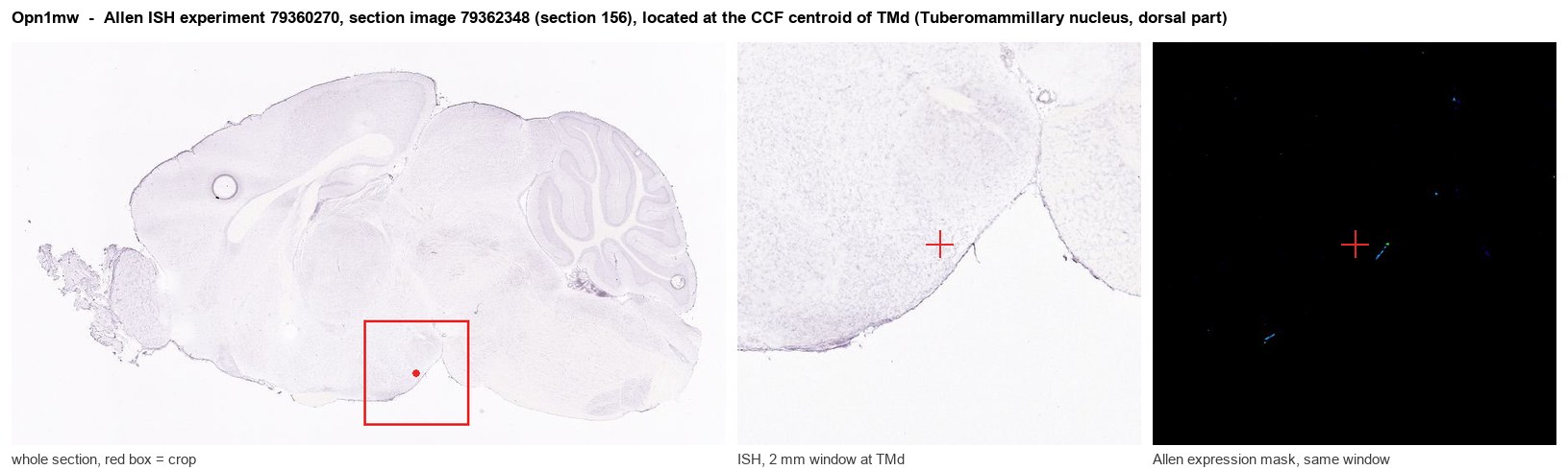
Allen ISH experiment 79360270, section image 79362348 (section 156): the section that contains the CCF centroid of Tuberomammillary nucleus, dorsal part according to the Allen image-to-atlas alignment (reference_to_image), with a 2 mm window around that point. Left: whole section, red box = window. Middle: ISH signal. Right: Allen's expression-mask view of the same window. open this image in the Allen viewer
Direct spatial evidence
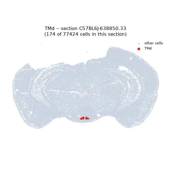
Regional expression figure
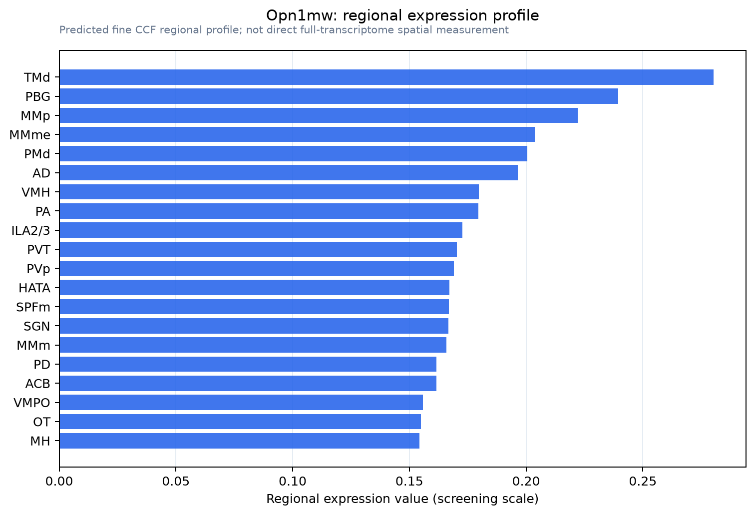
Predicted WMB × fine-CCF profile; not direct full-transcriptome spatial measurement.
Spatial profile
Evidence and specificity
- Evidence status
- PREDICTED_FINE
- Direct sources
- None; predicted localization only
- Exclusive threshold
- 12 positive regions out of 554
- Spatial specificity
- 0.277
- Top-region fraction
- 0.046
- Filter status
- PASS
Interpretation limits
- Cell-type-to-region projection is imputed expression, not direct spatial measurement.
- Adult reference atlases do not establish stability across age, sex, strain, state, or disease.
- Transcript abundance does not guarantee protein abundance or genetic-tool performance.