Mchr1
Melanin-concentrating hormone receptor 1 (MCH receptor 1) (MCH-R1) (MCHR-1) (G protein-coupled receptor 24) (MCH-1R) (MCH1R) (MCHR) (SLC-1) (Somatostatin receptor-like protein)
Spatial tier: REGION_BIASED · Class: GPCR · Top region: CA2sp (Field CA2, pyramidal layer) · Positive regions: 33/554
Function in CA2sp: evidence, prediction, innovation
- Gene function
- Mchr1 encodes melanin-concentrating hormone receptor 1, the only functional MCH receptor in rodents. It is a class-A GPCR coupling to Gi/Go and Gq, lowering cAMP and mobilising intracellular calcium in response to the hypothalamic peptide MCH, and it concentrates in the neuronal primary cilium. MCH-MCHR1 signalling regulates feeding, sleep and hippocampus-dependent learning.
- Region function
- CA2 stratum pyramidale is required for social recognition memory and is a major target of neuromodulatory peptides such as vasopressin and oxytocin that switch its output onto CA1 and lateral septum.
- Published in this region
- inferred No region-specific literature found. The closest work is in neighbouring CA1, where MCHR1 knockout mice show reduced AMPA and NMDA transmission, impaired LTP and LTD and passive-avoidance deficits (Pachoud 2010; Adamantidis 2005), and Diniz et al. 2020 mapped ciliary MCHR1 across the rodent hippocampus; Allen ISH in CA2sp is low (energy 0.65-0.86) and below CA1sp.
- Predicted function
- Prediction: ciliary MCHR1 on CA2 pyramidal neurons would act as a slow, state-dependent (sleep, arousal, satiety) modulator biasing CA2 excitability and plasticity rather than driving fast transmission. Intra-CA2 MCH should facilitate, and SNAP-94847 or CA2-restricted Mchr1 deletion should impair, social recognition memory and the NMDA-dependent component of CA2 synaptic drive.
- Innovation
- ★★★★☆ 4/5 MCH signalling in hippocampus has only been examined in CA1, leaving a plausible hypothalamic-to-CA2 state-gating pathway untested despite existing drugs and Cre lines.
- Tools
- Mchr1 knockout mice (Adamantidis 2005; Zhou 2005); MCHR1-CreER line (Engle 2018); antagonist SNAP-94847 with established in vivo dosing (David 2007); validated anti-MCHR1 antibody for ciliary labelling (Diniz 2020); CA2 access via Amigo2-Cre.
- References
Direct evidence located at the region
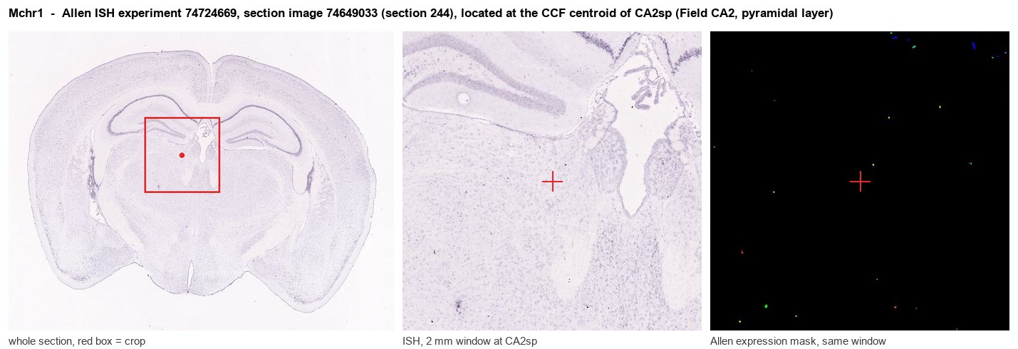
Allen ISH experiment 74724669, section image 74649033 (section 244): the section that contains the CCF centroid of Field CA2, pyramidal layer according to the Allen image-to-atlas alignment (reference_to_image), with a 2 mm window around that point. Left: whole section, red box = window. Middle: ISH signal. Right: Allen's expression-mask view of the same window. open this image in the Allen viewer
Direct spatial evidence
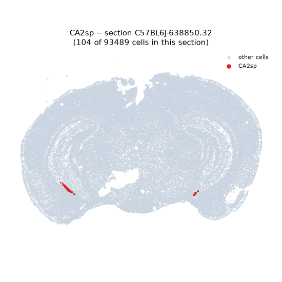
Regional expression figure
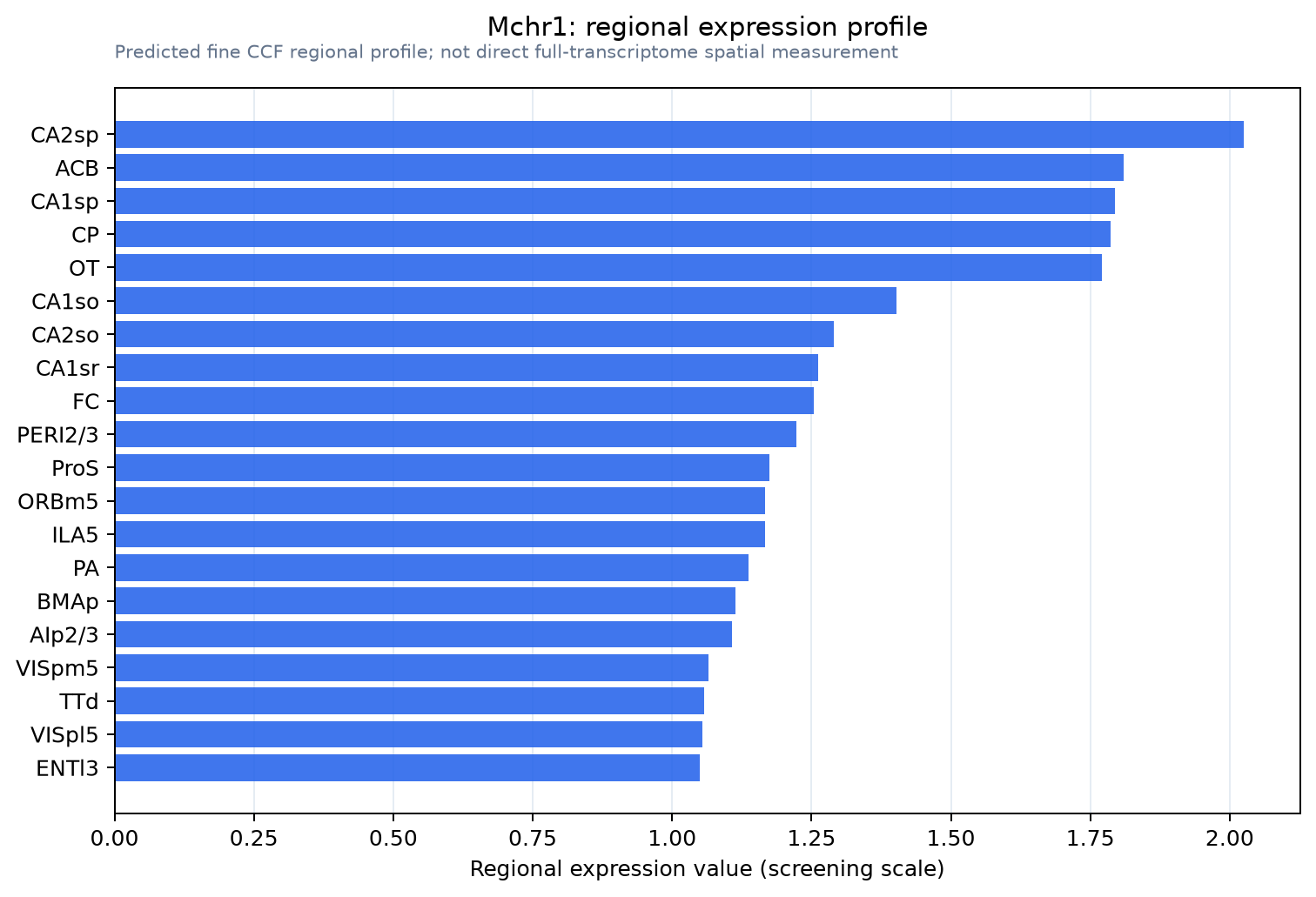
Predicted WMB × fine-CCF profile; not direct full-transcriptome spatial measurement.
Spatial profile
Evidence and specificity
- Evidence status
- PREDICTED_FINE
- Direct sources
- None; predicted localization only
- Exclusive threshold
- 12 positive regions out of 554
- Spatial specificity
- 0.284
- Top-region fraction
- 0.046
- Filter status
- PASS
Interpretation limits
- Cell-type-to-region projection is imputed expression, not direct spatial measurement.
- Adult reference atlases do not establish stability across age, sex, strain, state, or disease.
- Transcript abundance does not guarantee protein abundance or genetic-tool performance.