Gnrh1
Spatial tier: RESTRICTED · Class: OTHER · Top region: AVP (Anteroventral preoptic nucleus) · Positive regions: 28/554
43.5Discovery Score
35.0Targetability Score
CEvidence grade C
What this means: Gnrh1: fine CCF profile is restricted toward AVP (top regions: AVP, MEPO, AVPV, SO, ADP; 28 positive regions); direct spatial validation is still required.
Function in AVP: evidence, prediction, innovation
- Gene function
- Gnrh1 encodes preproGnRH, which is processed to the decapeptide gonadotropin-releasing hormone and a GnRH-associated peptide. GnRH is released episodically from median-eminence terminals into the hypophyseal portal blood, where it acts on the Gq-coupled GnRH receptor of pituitary gonadotropes to drive LH and FSH secretion. It is the apex signal of the hypothalamic-pituitary-gonadal axis.
- Region function
- The anteroventral preoptic/periventricular region is a strongly sexually dimorphic rostral preoptic nucleus whose kisspeptin and dopaminergic neurons form the female-specific luteinising-hormone surge generator and receive suprachiasmatic timing input for ovulation.
- Published in this region
- direct Direct. GnRH neurons in mouse form a loose continuum from the medial septum and diagonal band through the rostral preoptic area, including the anteroventral periventricular region, and Clarkson & Herbison 2006 showed that AVPV kisspeptin neurons are sexually dimorphic and project directly onto these GnRH cells. Herbison 2018 reviews how AVPV kisspeptin input drives the preovulatory GnRH surge, and Mason et al. 1986 showed that deletion of Gnrh1 produces the hypogonadal (hpg) mouse with infantile gonads and absent gonadotropins.
- Predicted function
- Prediction: remaining questions concern the local rather than pituitary actions of GnRH here - GnRH released from dendrites and collaterals within the AVPV is predicted to feed back onto kisspeptin and dopaminergic neurons to synchronise the surge. Selective deletion of Gnrh1 from rostral preoptic neurons while sparing more caudal populations, or local GnRH-receptor antagonism with cetrorelix, should desynchronise the LH surge and lengthen or disrupt the oestrous cycle without abolishing basal gonadotropin pulsatility.
- Innovation
- ★☆☆☆☆ 1/5 GnRH neurons in the preoptic region are among the most intensively studied neuroendocrine cells in the mouse brain.
- Tools
- Gnrh1-Cre and GnRH-GFP/GnRH-Pex5 reporter mice; hpg (Gnrh1 deletion) mice; GnRH agonists leuprolide and buserelin, antagonists cetrorelix and degarelix; LR-1 anti-GnRH antiserum; Kiss1-Cre and Th-Cre for the surrounding AVPV populations.
- References
Direct evidence located at the region
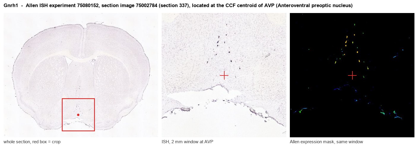
Allen ISH experiment 75080152, section image 75002784 (section 337): the section that contains the CCF centroid of Anteroventral preoptic nucleus according to the Allen image-to-atlas alignment (reference_to_image), with a 2 mm window around that point. Left: whole section, red box = window. Middle: ISH signal. Right: Allen's expression-mask view of the same window. open this image in the Allen viewer
Look it up yourself: Allen Mouse Brain ISH for Gnrh1Allen reference atlas: AVP (structure 263)ABC Atlas MERFISH viewer(in the ABC Atlas choose dataset MERFISH-C57BL6J-638850-CCF, colour cells by gene "Gnrh1" or by parcellation_substructure "AVP")All genes confined to AVP + 3-D brain
Direct spatial evidence
MERFISH REGION LOCATOR
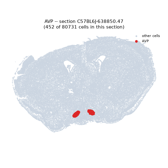
Regional expression figure
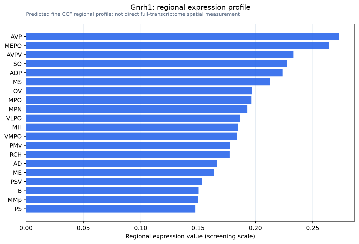
Predicted WMB × fine-CCF profile; not direct full-transcriptome spatial measurement.
Spatial profile
Evidence and specificity
- Evidence status
- PREDICTED_FINE
- Direct sources
- None; predicted localization only
- Exclusive threshold
- 12 positive regions out of 554
- Spatial specificity
- 0.321
- Top-region fraction
- 0.051
- Filter status
- PASS
Interpretation limits
- Cell-type-to-region projection is imputed expression, not direct spatial measurement.
- Adult reference atlases do not establish stability across age, sex, strain, state, or disease.
- Transcript abundance does not guarantee protein abundance or genetic-tool performance.