Drd5
D(1B) dopamine receptor (D(5) dopamine receptor) (Dopamine D5 receptor)
Spatial tier: RESTRICTED · Class: GPCR · Top region: PBG (Parabigeminal nucleus) · Positive regions: 25/554
44.6Discovery Score
40.2Targetability Score
CEvidence grade C
What this means: Drd5: fine CCF profile is restricted toward PBG (top regions: PBG, CA2sp, CA1sp, Xi, CA1so; 25 positive regions); direct spatial validation is still required.
Function in PBG: evidence, prediction, innovation
- Gene function
- Drd5 encodes the dopamine D5 receptor, a Gs-coupled D1-like GPCR stimulating adenylyl cyclase and cAMP/PKA signalling. It binds dopamine with roughly ten-fold higher affinity than D1 and is constitutively active, so it is thought to mediate tonic rather than phasic dopamine signalling, with a distribution distinct from D1.
- Region function
- The parabigeminal nucleus, the mammalian nucleus isthmi homologue, is a cholinergic and glutamatergic midbrain nucleus reciprocally connected with superior colliculus and projecting to central amygdala; it supports visual motion processing, attentional selection and looming-evoked defence.
- Published in this region
- inferred No region-specific literature found for D5 itself. Maltais et al. 2000 reported the parabigeminal nucleus as one of few rat regions with neurons double-labelled for D1 mRNA and D2 receptor protein, establishing that PBG neurons are dopaminoceptive. Ciliax et al. 2000 mapped D5 immunoreactivity broadly across midbrain and brainstem including superior colliculus neuropil, and Khan et al. 2000 found D5 on striatal cholinergic interneurons, the cell class PBG neurons most resemble.
- Predicted function
- Prediction: D5 in PBG cholinergic neurons provides high-affinity, partly constitutive Gs/cAMP drive setting spontaneous pacemaking rate and thereby scaling cholinergic gain in superficial superior colliculus. Since these neurons fire tonically via an I(A)-dependent mechanism (Goddard et al. 2007), D1-like agonism should accelerate firing, increase collicular acetylcholine release and lower escape thresholds, while Drd5 deletion or local antagonism should slow PBG firing and blunt SC-PBG-driven defensive responses.
- Innovation
- ★★★★★ 5/5 Dopaminergic modulation of the parabigeminal nucleus is unexplored even though the nucleus has a defined defensive circuit and ChAT-Cre access.
- Tools
- No D5-selective ligand exists, so combine D1-like agonist SKF-81297 and antagonist SCH-23390 with Drd5 knockout or conditional alleles (MGI/IMPC); D5 antibodies validated by Ciliax et al. 2000; PBG cholinergic neurons targetable with ChAT-IRES-Cre (JAX 006410) plus Cre-dependent AAV as in Tokuoka et al. 2020.
- References
Direct evidence located at the region
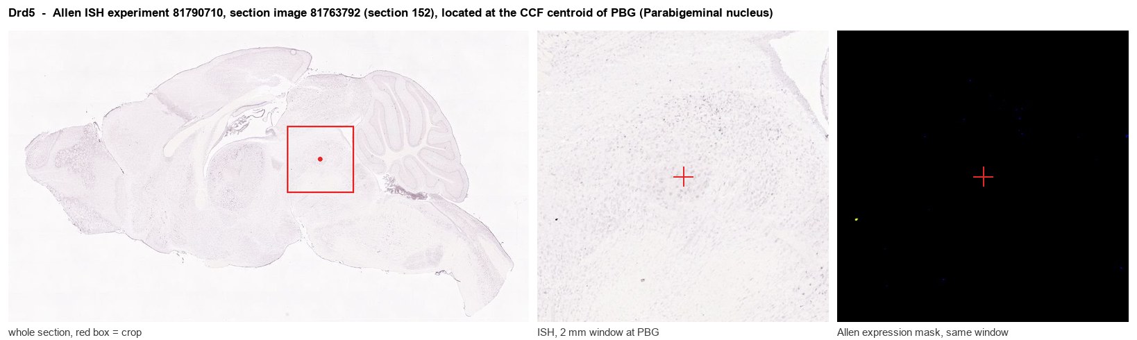
Allen ISH experiment 81790710, section image 81763792 (section 152): the section that contains the CCF centroid of Parabigeminal nucleus according to the Allen image-to-atlas alignment (reference_to_image), with a 2 mm window around that point. Left: whole section, red box = window. Middle: ISH signal. Right: Allen's expression-mask view of the same window. open this image in the Allen viewer
Look it up yourself: Allen Mouse Brain ISH for Drd5Allen reference atlas: PBG (structure 874)ABC Atlas MERFISH viewer(in the ABC Atlas choose dataset MERFISH-C57BL6J-638850-CCF, colour cells by gene "Drd5" or by parcellation_substructure "PBG")All genes confined to PBG + 3-D brain
Direct spatial evidence
MERFISH REGION LOCATOR
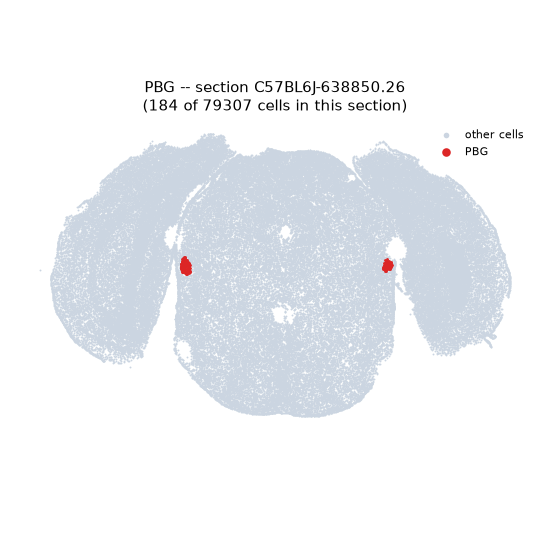
Regional expression figure
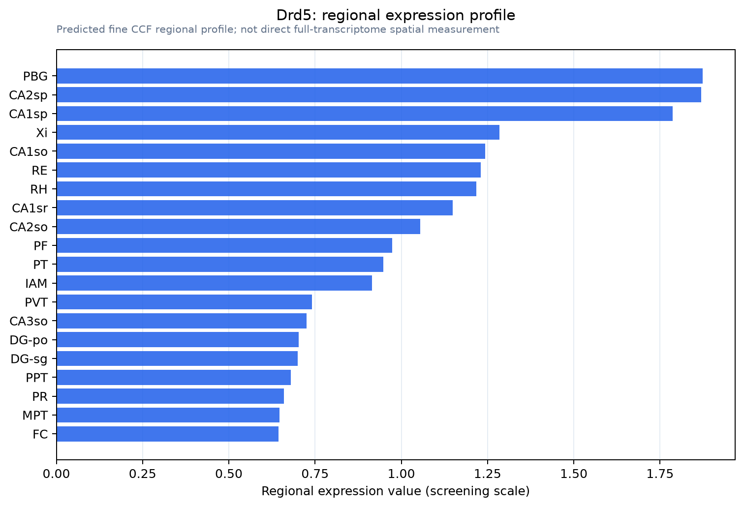
Predicted WMB × fine-CCF profile; not direct full-transcriptome spatial measurement.
Spatial profile
Evidence and specificity
- Evidence status
- PREDICTED_FINE
- Direct sources
- None; predicted localization only
- Exclusive threshold
- 12 positive regions out of 554
- Spatial specificity
- 0.324
- Top-region fraction
- 0.055
- Filter status
- PASS
Interpretation limits
- Cell-type-to-region projection is imputed expression, not direct spatial measurement.
- Adult reference atlases do not establish stability across age, sex, strain, state, or disease.
- Transcript abundance does not guarantee protein abundance or genetic-tool performance.