Cnr2
Cannabinoid receptor 2 (CB-2) (CB2) (mCB2)
Spatial tier: RESTRICTED · Class: GPCR · Top region: PCN (Paracentral nucleus) · Positive regions: 20/554
47.6Discovery Score
36.2Targetability Score
CEvidence grade C
What this means: Cnr2: fine CCF profile is restricted toward PCN (top regions: PCN, VPMpc, PO, MGd, VPM; 20 positive regions); direct spatial validation is still required.
Function in PCN: evidence, prediction, innovation
- Gene function
- Cnr2 encodes the CB2 cannabinoid receptor, a Gi/Go-coupled GPCR activated by 2-arachidonoylglycerol and anandamide that lowers cAMP and reduces neuronal firing and transmitter release. Long considered immune-restricted, it is now documented at low levels in brain microglia and neuron subsets, where it is strongly inducible by inflammation.
- Region function
- The paracentral nucleus is an intralaminar thalamic nucleus projecting diffusely to cortex and striatum; intralaminar nuclei support arousal, attentional selection, nociceptive-affective processing and thalamocortical interactions underlying conscious state.
- Published in this region
- inferred No region-specific literature found. Grabon et al. 2024 used cell sorting, calibrated RT-qPCR and single-nucleus RNA-seq to show CB2 mRNA is present at low levels in every mouse brain region examined, carried mainly by a minority of microglia and a smaller fraction of neurons, with inflammation-dependent regulation. Zhang et al. 2021 demonstrated genuine neuronal CB2 in red nucleus glutamate neurons, where JWH133 slowed firing in wild-type but not CB2-knockout mice.
- Predicted function
- Prediction: PCN CB2 is most likely carried by microglia plus a sparse population of thalamocortical glutamate neurons, where Gi coupling would dampen firing and thalamocortical drive. Extending the red nucleus result, local JWH133 should reduce PCN firing and lower cortical arousal, attentional gain and affective pain responses, while Cnr2 deletion or AM630 should increase them, with effects amplified after inflammatory challenge. Cell-type attribution must be settled first, since microglial CB2 could produce the same readout indirectly.
- Innovation
- ★★★★☆ 4/5 Neuronal CB2 has been validated in only a handful of nuclei and never in intralaminar thalamus, a region central to arousal and pain.
- Tools
- Agonists JWH133 and HU-308, antagonists AM630 and SR144528; Cnr2 global and conditional knockouts as the essential specificity control; a CB2-KO-eGFP reporter line described by Xi and colleagues; thalamic neurons targetable with Slc17a6-Cre, microglia with Cx3cr1-CreER.
- References
Direct evidence located at the region
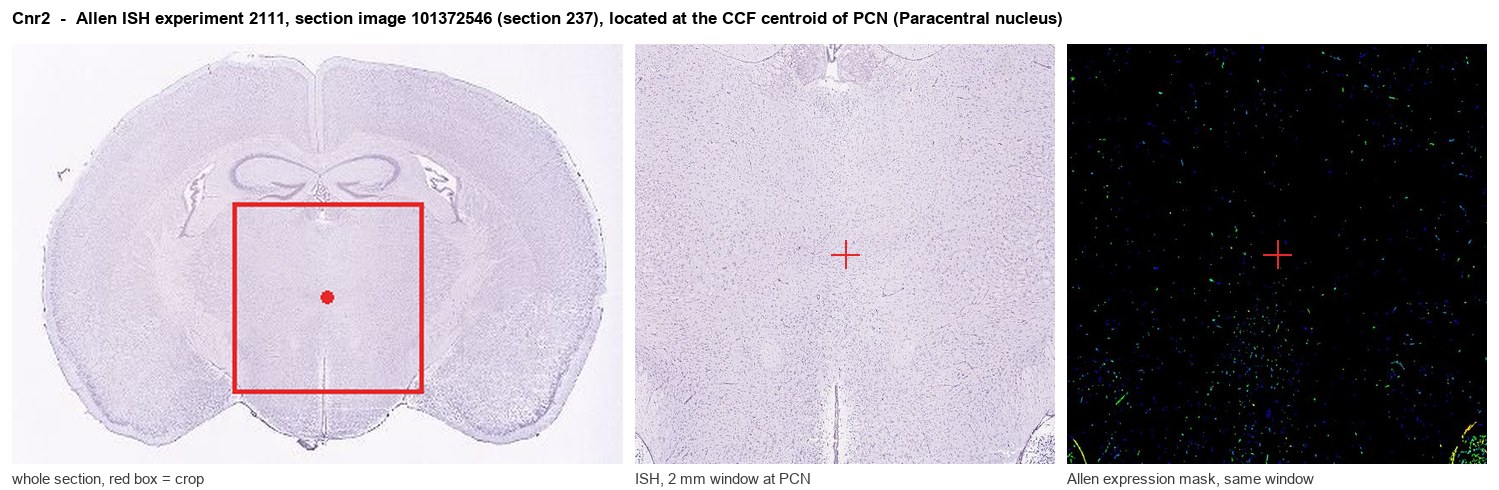
Allen ISH experiment 2111, section image 101372546 (section 237): the section that contains the CCF centroid of Paracentral nucleus according to the Allen image-to-atlas alignment (reference_to_image), with a 2 mm window around that point. Left: whole section, red box = window. Middle: ISH signal. Right: Allen's expression-mask view of the same window. open this image in the Allen viewer
Look it up yourself: Allen Mouse Brain ISH for Cnr2Allen reference atlas: PCN (structure 907)ABC Atlas MERFISH viewer(in the ABC Atlas choose dataset MERFISH-C57BL6J-638850-CCF, colour cells by gene "Cnr2" or by parcellation_substructure "PCN")All genes confined to PCN + 3-D brain
Direct spatial evidence
MERFISH REGION LOCATOR
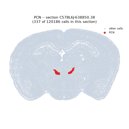
Regional expression figure
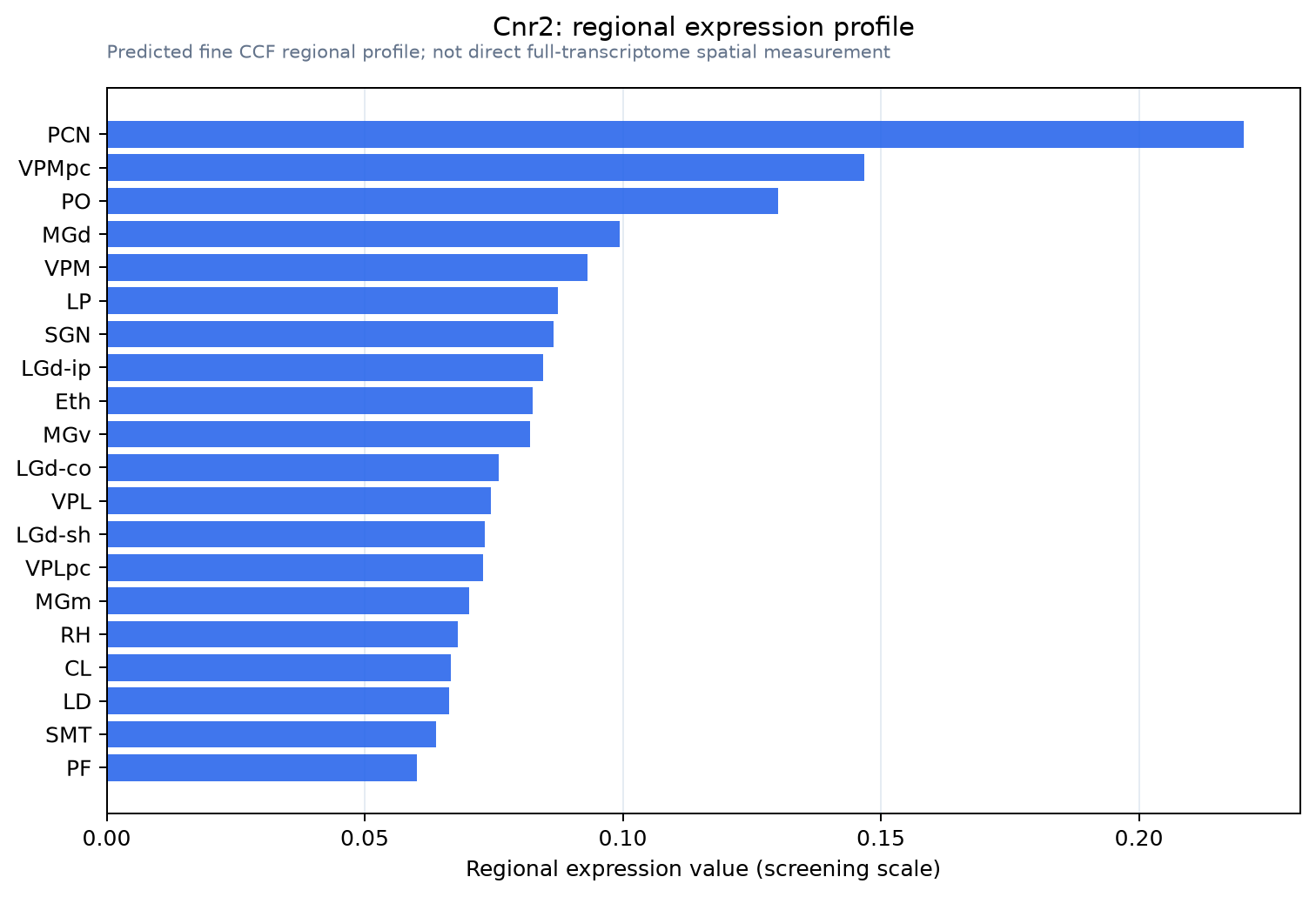
Predicted WMB × fine-CCF profile; not direct full-transcriptome spatial measurement.
Spatial profile
Evidence and specificity
- Evidence status
- PREDICTED_FINE
- Direct sources
- None; predicted localization only
- Exclusive threshold
- 12 positive regions out of 554
- Spatial specificity
- 0.349
- Top-region fraction
- 0.088
- Filter status
- PASS
Interpretation limits
- Cell-type-to-region projection is imputed expression, not direct spatial measurement.
- Adult reference atlases do not establish stability across age, sex, strain, state, or disease.
- Transcript abundance does not guarantee protein abundance or genetic-tool performance.