Best1
Bestrophin-1 (Vitelliform macular dystrophy protein 2 homolog)
Spatial tier: REGION_BIASED · Class: ION_CHANNEL · Top region: MEPO (Median preoptic nucleus) · Positive regions: 32/554
43.3Discovery Score
38.1Targetability Score
CEvidence grade C
What this means: Best1: fine CCF profile is region-biased toward MEPO (top regions: MEPO, OV, TMd, AVPV, ME); this is a discovery lead, not direct proof.
Function in MEPO: evidence, prediction, innovation
- Gene function
- Best1 encodes bestrophin-1, a calcium-activated anion channel whose human mutations cause Best vitelliform macular dystrophy. In brain it is predominantly astrocytic and unusually permeable not only to chloride but to glutamate and GABA, making it a channel-mediated gliotransmitter release route. It supplies the tonic GABA that activates extrasynaptic GABA-A receptors, and reactive astrocytes redistribute it to release excess GABA.
- Region function
- The median preoptic nucleus is the integrative node of the lamina terminalis, combining osmotic, angiotensin and baroreceptor signals to drive thirst and vasopressin release, and also controlling thermoregulatory heat loss, sleep initiation and cardiovascular set-point.
- Published in this region
- inferred No region-specific literature found. The closest evidence is Pandit 2015, showing astroglial GABA uptake and tonic GABA-A inhibition regulate presympathetic hypothalamic paraventricular neurons, and Kwak 2020, showing astrocytic Best1-mediated tonic GABA sets sensory acuity in thalamus; Allen ISH gives MEPO 0.088 versus 0.126 for grey matter (experiment 70302992), consistent with diffuse astrocytic expression.
- Predicted function
- Prediction: Best1 in MEPO is an astrocytic tonic GABA conduit clamping the excitability of glutamatergic thirst- and heat-loss-promoting neurons, setting the threshold at which osmotic and thermal drive becomes drinking or vasomotor heat loss. Astrocyte-specific deletion should lower that threshold, giving exaggerated drinking to modest osmotic challenge, blunted febrile responses or fragmented NREM sleep; overexpression should dampen them.
- Innovation
- ★★★★☆ 4/5 Astrocytic tonic GABA is mapped in hippocampus, cerebellum and thalamus, never in the lamina terminalis.
- Tools
- Best1 floxed crossed to hGFAP-CreERT2 for astrocyte-specific deletion (Joo 2024); Best1 knockout with astrocytic rescue (Pandit 2020); pSico-Red Best1 shRNA AAV (Jung 2016); siBest1 nanoparticles (Xiong 2025); blockers NPPB, CaCCinh-A01.
- References
Direct evidence located at the region
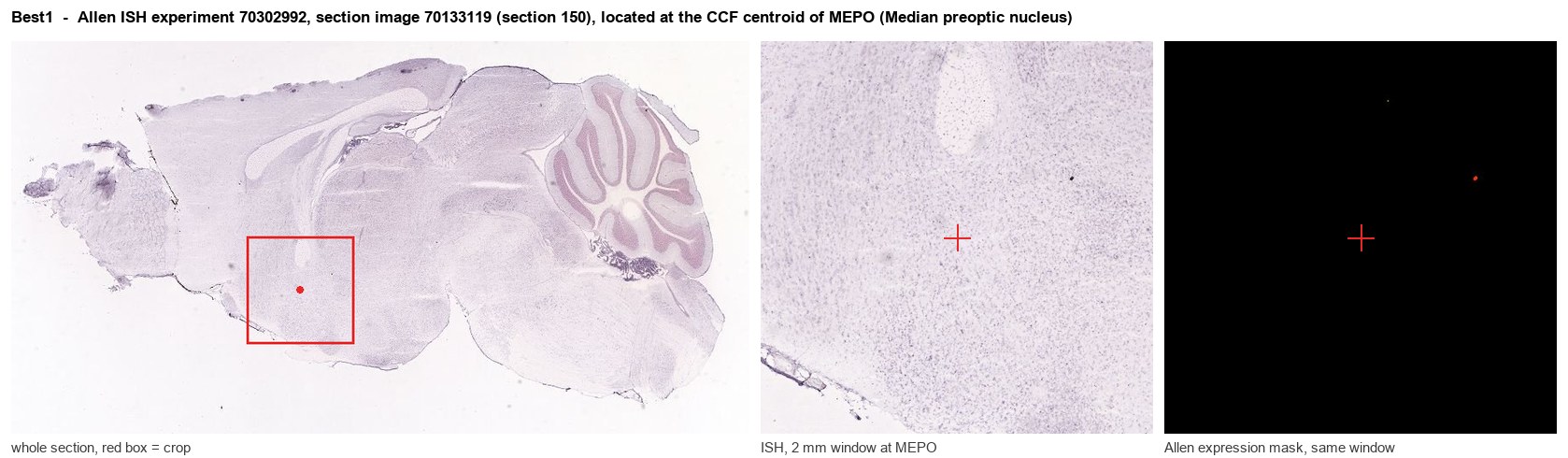
Allen ISH experiment 70302992, section image 70133119 (section 150): the section that contains the CCF centroid of Median preoptic nucleus according to the Allen image-to-atlas alignment (reference_to_image), with a 2 mm window around that point. Left: whole section, red box = window. Middle: ISH signal. Right: Allen's expression-mask view of the same window. open this image in the Allen viewer
Look it up yourself: Allen Mouse Brain ISH for Best1Allen reference atlas: MEPO (structure 452)ABC Atlas MERFISH viewer(in the ABC Atlas choose dataset MERFISH-C57BL6J-638850-CCF, colour cells by gene "Best1" or by parcellation_substructure "MEPO")All genes confined to MEPO + 3-D brain
Direct spatial evidence
MERFISH REGION LOCATOR
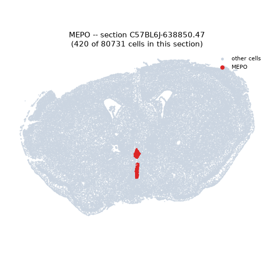
Regional expression figure
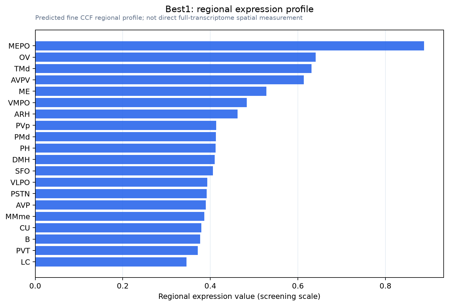
Predicted WMB × fine-CCF profile; not direct full-transcriptome spatial measurement.
Spatial profile
Evidence and specificity
- Evidence status
- PREDICTED_FINE
- Direct sources
- None; predicted localization only
- Exclusive threshold
- 12 positive regions out of 554
- Spatial specificity
- 0.315
- Top-region fraction
- 0.055
- Filter status
- PASS
Interpretation limits
- Cell-type-to-region projection is imputed expression, not direct spatial measurement.
- Adult reference atlases do not establish stability across age, sex, strain, state, or disease.
- Transcript abundance does not guarantee protein abundance or genetic-tool performance.