Avpr1b
Vasopressin V1b receptor (V1bR) (AVPR V1b) (AVPR V3) (Antidiuretic hormone receptor 1b) (Vasopressin V3 receptor)
Spatial tier: STRICT_EXCLUSIVE · Class: GPCR · Top region: CA2sp (Field CA2, pyramidal layer) · Positive regions: 4/554
54.3Discovery Score
44.8Targetability Score
CEvidence grade C
What this means: Avpr1b: predicted fine-region profile is strict-exclusive, with 4 positive regions out of 554 (limit 12); direct spatial validation is required.
Function in CA2sp: evidence, prediction, innovation
- Gene function
- Avpr1b encodes the vasopressin V1b receptor, a Gq-coupled GPCR that signals through phospholipase C, IP3 and calcium. It was first characterised on pituitary corticotrophs, where vasopressin potentiates CRH-driven ACTH release; in the brain its expression is narrow and strikingly enriched in hippocampal CA2 pyramidal neurons.
- Region function
- The CA2 pyramidal layer receives dense supramammillary and hypothalamic vasopressin/oxytocin input and is required for social recognition memory and for normal social aggression, relaying CA3 and entorhinal input to CA1.
- Published in this region
- direct Pagani et al. 2015 (Mol Psychiatry) showed Avpr1b mRNA is concentrated in dorsal CA2 pyramidal cells, that knockouts show reduced social (not predatory) aggression in both sexes, and that lentiviral Avpr1b re-expression in dorsal CA2 rescued aggression; V1b agonist enhanced CA2 synaptic responses through an NMDA-receptor-dependent mechanism. Williams Avram et al. 2019 built an Avpr1b-Cre knock-in labelling dorsal and ventral CA2. Hitti and Siegelbaum 2014 established CA2 as necessary for social memory.
- Predicted function
- Prediction: vasopressin acting on V1b in CA2 pyramidal cells raises excitability and synaptic gain, gating whether a social encounter is encoded and whether it triggers aggression. Knockout or CA2-targeted antagonism (SSR149415) should reduce social aggression and weaken social recognition; chemogenetic activation of Avpr1b-Cre CA2 neurons should promote social investigation and aggressive responding.
- Innovation
- ★☆☆☆☆ 1/5 Avpr1b in CA2 is a textbook example with a dedicated Cre line, knockout rescue experiments and selective antagonists.
- Tools
- Avpr1b-Cre knock-in (Williams Avram et al. 2019), Avpr1b knockout mice, Amigo2-Cre for CA2 pyramidal cells, V1b antagonist SSR149415 (nelivaptan), selective V1b agonist d[Leu4,Lys8]VP, lentiviral/AAV Avpr1b rescue vectors.
- References
Direct evidence located at the region
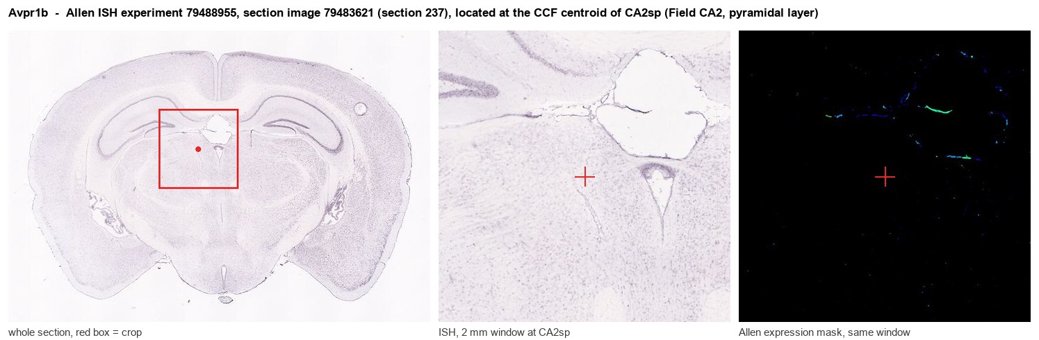
Allen ISH experiment 79488955, section image 79483621 (section 237): the section that contains the CCF centroid of Field CA2, pyramidal layer according to the Allen image-to-atlas alignment (reference_to_image), with a 2 mm window around that point. Left: whole section, red box = window. Middle: ISH signal. Right: Allen's expression-mask view of the same window. open this image in the Allen viewer
Look it up yourself: Allen Mouse Brain ISH for Avpr1bAllen reference atlas: CA2sp (structure 446)ABC Atlas MERFISH viewer(in the ABC Atlas choose dataset MERFISH-C57BL6J-638850-CCF, colour cells by gene "Avpr1b" or by parcellation_substructure "CA2sp")All genes confined to CA2sp + 3-D brain
Direct spatial evidence
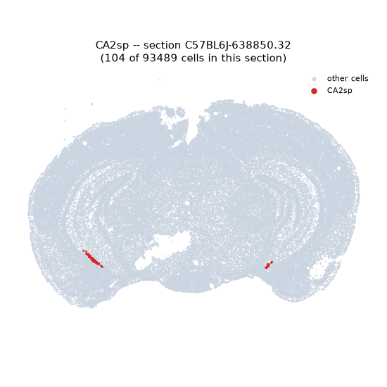
MERFISH REGION LOCATOR
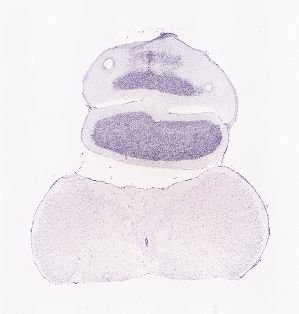
DIRECT ALLEN ISH
DIRECT ALLEN ISH
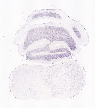
Regional expression figure
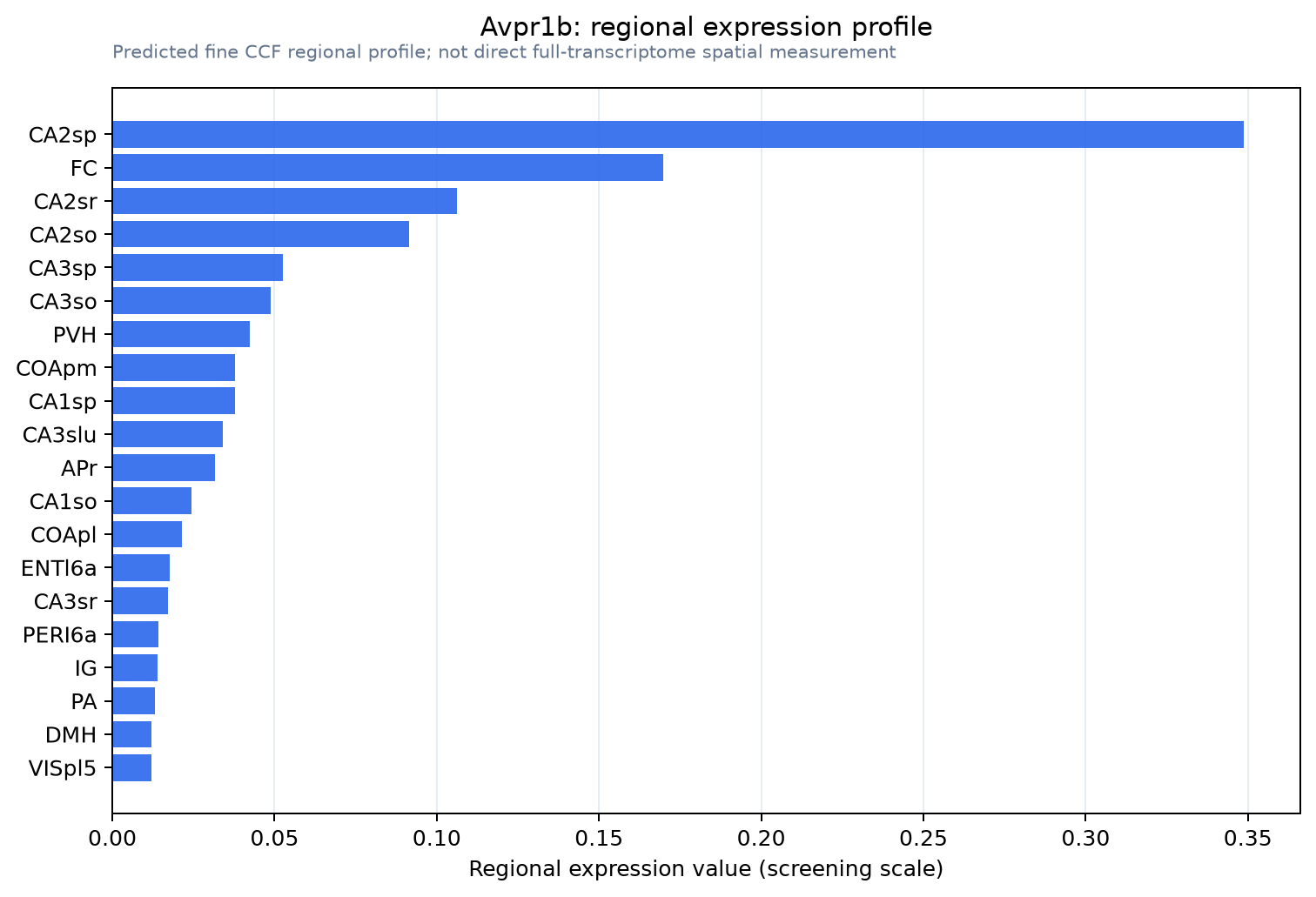
Predicted WMB × fine-CCF profile; not direct full-transcriptome spatial measurement.
Spatial profile
Evidence and specificity
- Evidence status
- PREDICTED_FINE
- Direct sources
- None; predicted localization only
- Exclusive threshold
- 12 positive regions out of 554
- Spatial specificity
- 0.496
- Top-region fraction
- 0.258
- Filter status
- PASS
Interpretation limits
- Cell-type-to-region projection is imputed expression, not direct spatial measurement.
- Adult reference atlases do not establish stability across age, sex, strain, state, or disease.
- Transcript abundance does not guarantee protein abundance or genetic-tool performance.