Adgre1
Adhesion G protein-coupled receptor E1 (Cell surface glycoprotein F4/80) (EGF-like module receptor 1) (EGF-like module-containing mucin-like hormone receptor-like 1) (EMR1 hormone receptor)
Spatial tier: RESTRICTED · Class: GPCR · Top region: LC (Locus ceruleus) · Positive regions: 13/554
47.3Discovery Score
38.1Targetability Score
CEvidence grade C
What this means: Adgre1: fine CCF profile is restricted toward LC (top regions: LC, SLC, DMX, AId1, TEa1; 13 positive regions); direct spatial validation is still required.
Function in LC: evidence, prediction, innovation
- Gene function
- ADGRE1 encodes EMR1, better known in mouse as F4/80. It is an adhesion-class G-protein-coupled receptor with a string of extracellular EGF-like repeats and a GAIN domain. F4/80 is the classic pan-macrophage and microglia surface marker in mouse and is required for the induction of peripheral immune tolerance, while the human orthologue EMR1 is restricted to eosinophils; no ligand or downstream signalling output is firmly established for either.
- Region function
- The locus coeruleus is the principal noradrenergic nucleus of the brain; its small population of neurons innervates nearly the whole forebrain and sets arousal, attention, stress reactivity, sleep-wake transitions and network gain.
- Published in this region
- inferred No region-specific literature found. Adgre1/F4/80 in brain labels microglia and perivascular and meningeal macrophages rather than neurons (Hamann et al. 2007), and Wasinski et al. 2023 used hypothalamic Adgre1 as a neuroinflammatory marker; a locus coeruleus signal therefore most plausibly reflects the nucleus's dense perivascular macrophage and microglial population rather than noradrenergic neurons themselves.
- Predicted function
- Prediction: ADGRE1-positive macrophages and microglia in the locus coeruleus are predicted to act as local immune sentinels whose activation state gates noradrenergic excitability, providing a route by which systemic inflammation produces sickness-associated changes in arousal and attention. Microglia-restricted Adgre1 deletion or macrophage depletion with clodronate or CSF1R inhibitors in LC is predicted to leave baseline sleep-wake architecture intact but to blunt the LPS-induced rise in LC firing and the accompanying sickness-related hypersomnolence and reduced exploratory behaviour; F4/80 immunolabelling combined with Dbh-Cre photometry is the natural first experiment.
- Innovation
- ★★★☆☆ 3/5 Neuroimmune modulation of the locus coeruleus is an interesting and under-explored idea, but because Adgre1 almost certainly marks glia rather than LC neurons the finding is less surprising and the reagents are standard immunology tools.
- Tools
- F4/80 (Adgre1)-Cre and Adgre1-knockout mice; anti-F4/80 antibody clones BM8 and CI:A3-1; Cx3cr1-CreER and Lyve1-Cre for microglia versus perivascular macrophages; clodronate liposomes and CSF1R inhibitors PLX5622/PLX3397 for depletion; Dbh-Cre and Net-Cre plus GRAB-NE for the noradrenergic readout.
- References
Direct evidence located at the region
No Allen ISH section could be located at this region's centroid for the recorded experiments.
Look it up yourself: Allen Mouse Brain ISH for Adgre1Allen reference atlas: LC (structure 147)ABC Atlas MERFISH viewer(in the ABC Atlas choose dataset MERFISH-C57BL6J-638850-CCF, colour cells by gene "Adgre1" or by parcellation_substructure "LC")All genes confined to LC + 3-D brain
Direct spatial evidence
MERFISH REGION LOCATOR
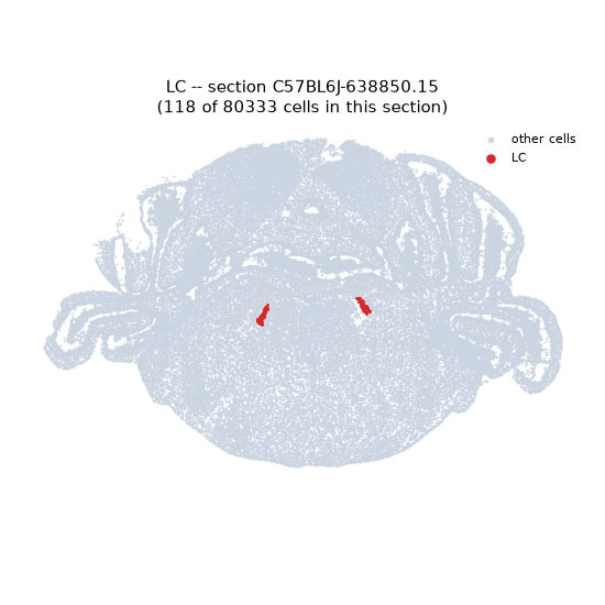
Regional expression figure
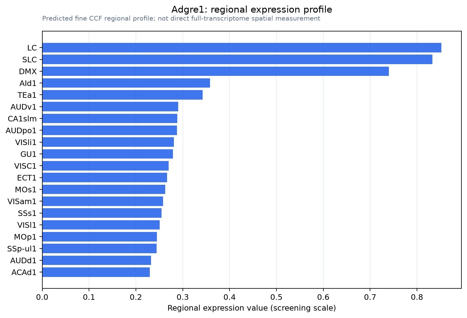
Predicted WMB × fine-CCF profile; not direct full-transcriptome spatial measurement.
Spatial profile
Evidence and specificity
- Evidence status
- PREDICTED_FINE
- Direct sources
- None; predicted localization only
- Exclusive threshold
- 12 positive regions out of 554
- Spatial specificity
- 0.323
- Top-region fraction
- 0.077
- Filter status
- PASS
Interpretation limits
- Cell-type-to-region projection is imputed expression, not direct spatial measurement.
- Adult reference atlases do not establish stability across age, sex, strain, state, or disease.
- Transcript abundance does not guarantee protein abundance or genetic-tool performance.